The Norwood Scale Explained: What Your Hair Loss Stage Really Means

Updated: Jun 2

When men first notice a shift in their hairline or a loss of volume at the crown, the immediate reaction is to search for a baseline. How bad is it? How far will it go? Am I a candidate for a hair transplant? In the realm of clinical hair restoration, we do not rely on subjective descriptions. We rely on the Norwood Scale (originally the Hamilton-Norwood Scale)—the universal medical standard used to classify, diagnose, and track the progression of male pattern baldness (androgenetic alopecia).
Understanding the Norwood Scale is not just about labeling your current hair loss. It is about architectural forecasting. By identifying your current stage, a master clinical team can predict your future loss trajectory, calculate your required graft counts, and design a lifelong surgical blueprint.
Here is Eva Estetica’s comprehensive clinical breakdown of the 7 stages of the Norwood Scale, what they biologically represent, and when surgical intervention becomes necessary.
Table of Contents: The Norwood Scale Explained
The 7 Stages of the Norwood Scale: A Clinical Breakdown

The Norwood Scale tracks the highly predictable, geometric retreat of DHT-sensitive hair follicles throughout the stages of male pattern baldness. It primarily monitors two specific anatomical zones: the frontal temporal peaks (the hairline) and the vertex (the crown).
Stage 1: The Juvenile Hairline
Visual Presentation: A full, dense head of hair with a flat, relatively straight frontal hairline.
The Biological Reality: This is the baseline hairline of your youth. There is zero evidence of DHT-induced follicle miniaturization, shedding, or genetic recession.
Clinical Action: No receding hairline treatment, medical therapy, or surgical intervention is required or recommended.
Stage 2: The Mature Hairline
Visual Presentation: The hairline experiences a very slight, symmetrical recession at the temples, usually receding by about 1 to 1.5 centimeters above the upper brow crease.
The Biological Reality: Patients often panic at this stage, mistaking it for active hair loss. However, this is not clinical baldness. This is the natural, biological transition from a teenage hairline to a mature, adult male facial frame.
Clinical Action: An elite surgeon will virtually always refuse to perform a hair transplant on a Stage 2 hairline. Surgically filling in these minor corners disrupts natural facial aging, creates aesthetic friction in your 40s and 50s, and exhausts your finite donor grafts unnecessarily.
Stage 3 & Stage 3-Vertex: The Tipping Point
Visual Presentation: The recession at the temples deepens significantly, forming a distinct "M", "V", or "U" shape that becomes difficult to camouflage with styling. In the Stage 3-Vertex variant, the frontal recession is accompanied by a noticeable bald spot or severe crown thinning at the back of the head.
The Biological Reality: This is the critical tipping point and the first stage universally classified as clinical baldness. The DHT hormone has successfully miniaturized the vulnerable follicles in these distinct, localized zones.
Clinical Action: This is the ideal stage for early intervention. Medical therapy (like Finasteride or Minoxidil) is heavily utilized here to stabilize the native hair, while a precision Norwood 3 hair transplant can flawlessly reconstruct the frontal boundary before the loss progresses further.
Stage 4: The Bridge
Visual Presentation: The frontal recession deepens further, and the bald spot on the crown expands outward. However, there is still a distinct, moderately dense "bridge" of healthy hair separating the front of the head from the crown.
The Biological Reality: Hair loss progression is accelerating. The genetically resistant hairs forming the "bridge" are now flanked by aggressive miniaturization on both sides, making the scalp highly visible under direct light.
Clinical Action: Surgical restoration is highly effective here, but it requires strategic architectural planning. The surgeon must evaluate your donor capacity and decide whether to prioritize restoring the frontal third to reframe the face, or carefully distributing grafts to provide lighter coverage across both zones.
Stage 5: The Thinning Bridge
Visual Presentation: The balding areas at the front and the crown continue to enlarge significantly. The dense "bridge" of hair separating them begins to severely narrow, thin, and break down into isolated hairs.
The Biological Reality: This marks the shift into advanced male pattern baldness. The DHT sensitivity is spreading across the entire mid-scalp, effectively dismantling the last barrier between the front and the back.
Clinical Action: Achieving high-density restoration at Stage 5 requires elite donor area management. Non-surgical medication is strongly recommended to preserve whatever remains of the mid-scalp bridge, while a high-graft FUE transplant focuses on rebuilding the most cosmetically important areas.
Stage 6: The Merge
Visual Presentation: The bridge of hair vanishes entirely. The frontal baldness and the crown baldness merge into one single, massive expanse of bare skin across the top of the head.
The Biological Reality: The vast majority of the upper scalp has succumbed to genetic hair loss, leaving only the hair on the sides and the back of the head.
Clinical Action: Because the recipient area is now vastly larger than the available donor area, full, dense coverage in a single surgery is usually biologically impossible. Restoration at Stage 6 often requires a multi-stage approach, prioritizing a conservative, age-appropriate hairline design to restore the facial profile while leaving the crown intentionally lighter.
Stage 7: The Horseshoe
Visual Presentation: The most severe stage of hair loss progression. All hair on the top of the head is gone, leaving only a thin, horseshoe-shaped band of hair around the sides and lower back of the head.
The Biological Reality: Only the most profoundly DHT-resistant follicles located in the strictest parameters of the "safe donor zone" remain alive.
Clinical Action: Candidacy for a hair transplant at Stage 7 is exceptionally rare and highly dependent on having unusually dense, thick donor hair. Even when approved for surgery, patient expectations must be strictly managed—the goal is framing the face with light coverage, as a full head of hair cannot be biologically restored.
Deep Dive: A successful restoration is built on understanding the exact differences between localized recession and widespread shedding. Discover how we navigate complex hair loss patterns in our diagnostic guide: Diffuse Thinning vs Receding Hairline: Why the Difference Matters.
The Speed of Progression: How Fast Will You Lose It?

Once a patient identifies their current Norwood stage, the immediate follow-up question is invariably: "How long do I have until I reach the next stage?" It is human nature to want a strict timeline or a mathematical formula to predict exactly when you will become a Stage 4 or Stage 5. However, there is a common misconception that male pattern baldness is a slow, steady, and perfectly linear decline.
In clinical reality, androgenetic alopecia (AGA) is uniquely individualized and progresses in a highly unpredictable pattern.
The Unpredictable Nature of Miniaturization
You cannot accurately predict hair loss progression using a calendar. Rather than a constant, daily shedding of a set number of hairs, DHT-sensitive follicles miniaturize over time in a variable pattern.
Most patients experience periods of apparent stability—where their hairline seems unchanged for years—followed by periods of highly visible change and accelerated thinning. This occurs because DHT (Dihydrotestosterone) disrupts the normal hair growth cycle. It forces the active growth phase (anagen) to become progressively shorter, meaning hairs return microscopically thinner and weaker with each cycle until the follicle eventually stops producing hair entirely.
Altering the Trajectory: The Role of Medical Therapy
While genetic hair loss is progressive, its trajectory can be significantly altered with proper medical management. A master surgical plan often relies on these therapies to protect the native hair sitting directly behind a newly transplanted hairline. However, it is vital to understand the precise roles and limitations of these treatments:
Finasteride (DHT Blockers): This oral or topical medication addresses the biological root cause by inhibiting the conversion of testosterone into DHT. Its primary clinical function is to slow down or completely halt further hair loss.
Minoxidil (Growth Stimulants): Operating through a different mechanism, Minoxidil helps support new hair growth and maintains follicle health by extending the active growth phase and increasing blood flow to the scalp.
The Maintenance Caveat: Neither of these treatments is a permanent cure. The benefits of both Finasteride and Minoxidil rely entirely on continuous, ongoing use. If you stop taking the medication, the protective benefits will fade, and your hair loss will eventually resume its natural genetic course.
Understanding the Side Effects of DHT Blockers
At Eva Estetica, clinical honesty is the foundation of our practice. While DHT blockers like Finasteride are the most highly effective tools for preserving your biological hair, they are systemic medical treatments and must be approached with informed consent.
Because Finasteride alters your hormone profile by reducing DHT, a small percentage of patients may experience systemic side effects. These can include decreased libido, erectile dysfunction, or, in rare cases, mood changes. For the vast majority of men, the medication is well-tolerated, and side effects resolve if the dosage is adjusted or the medication is discontinued.
Because every patient's biological tolerance is different, determining your surgical and medical roadmap requires a comprehensive consultation. An ethical clinic will always weigh the cosmetic benefits of DHT blockers against your overall physiological well-being before writing a prescription.
Deep Dive: Before you can accurately track your stage on the chart, it is crucial to understand the biological engine driving the retreat. Discover exactly how your genetics and Dihydrotestosterone (DHT) trigger follicle miniaturization in our foundational guide: Male Pattern Baldness Explained: Genetics, DHT, and Hair Loss Planning.
Beyond the Standard Scale: The Norwood Variants

While the standard 1–7 progression maps the most common pattern of male pattern baldness—an "M" shape forming at the front and eventually merging with a bald crown—hair loss is rarely a one-size-fits-all condition. When Dr. O'Tar Norwood revised this scale, he identified several distinct, alternative trajectories.
Understanding these variants is crucial because they fundamentally change your timeline for intervention and dictate an entirely different surgical strategy.
1. The "Class A" Variant: The Unconventional Trajectory
While less common than the standard pattern, a notable subset of men experience the Norwood Class A progression. In this variant, the hair does not form the classic deep temporal angles (the "M" or "V" shape), nor does it leave an isolated island of hair in the mid-scalp. Furthermore, these patients typically do not develop an isolated bald spot on the crown early on.
Instead, the entire frontal hairline marches backward in a uniform, relatively straight line—moving like a receding tide across the scalp. The Class A variant is tracked in its own sub-stages (Stage 2A, 3A, 4A, and 5A) depending on how far back the hairline has traveled.
Surgical Advantage: Because Class A patients do not have to worry about a rapidly expanding bald spot on their crown, their surgical planning is often more straightforward. The surgeon does not have to split the donor grafts between the front and the back of the head, allowing the biological capital to be dedicated entirely to rebuilding a single, dense frontal zone.
2. The Vertex Variant (e.g., Stage 3-Vertex)
The Vertex Variant is characterized by aggressive, early-onset hair loss specifically localized at the crown of the head (the vertex). A patient might only have mild to moderate frontal recession (presenting as a standard Norwood Stage 3), but simultaneously suffer from a distinct, highly visible bald spot on the back of the head.
Surgical Strategy: The crown is one of the most challenging areas in hair restoration. Because of the whorl (the spiral angle of the hair growth), this area requires a high volume of grafts to create the illusion of density. For Vertex variants, an elite clinic will often prioritize non-surgical medical stabilization (like Finasteride or Minoxidil) to thicken the crown before attempting to surgically pack it with precious donor grafts.
3. The Diffuse Variants: DPA vs. DUPA
While the traditional Norwood scale measures areas of slick, bare skin, the Diffuse Variant tracks a generalized loss of volume. In clinical practice, distinguishing the type of diffuse loss is incredibly important because the stability of the donor area is what makes transplantation possible in the first place.
Diffuse Patterned Alopecia (DPA): In DPA, the hairline boundaries might stay anchored at a Norwood Stage 2 or 3, but the overall density across the top of the scalp drops uniformly. Crucially, the donor area remains stable. While DPA cases can sometimes be treated surgically, they require far more careful planning than a bare, receding hairline. Because the balding area is still filled with weak native hairs, the surgeon must work meticulously to prevent surgical shock loss. Techniques like Direct Hair Implantation (DHI) can be highly useful when working precisely between existing hairs, though other advanced implantation methods can also be effective.
Diffuse Unpatterned Alopecia (DUPA): DUPA involves diffuse, unpatterned thinning that extends into the traditional donor area at the back and sides of the scalp. Diffuse donor miniaturization is a major clinical warning sign. Because the donor hair itself lacks long-term genetic stability, it cannot be safely relocated. This instability often makes DUPA patients a poor or unsafe candidate for surgical intervention, requiring a pivot to medical management instead.
Deep Dive: A common misconception is that you should rush into surgery the moment you notice hair loss. Discover why we strictly refuse to operate on Stage 1 and Stage 2 hairlines in our clinical guide on when to get a hair transplant and why timing is everything.
The Surgical Mathematics: Graft Estimations by Norwood Stage

One of the most frequent questions we receive during consultations is: "How many grafts do I need for my Norwood stage?" While patients naturally look for a definitive number, it is crucial to understand that graft counts are highly variable. Two patients at the exact same Norwood stage might require vastly different graft counts depending on their individual anatomy, head size, hair caliber (thickness), and precise donor density.
While absolute numbers can only be finalized through a digital microscopic audit, we can provide general architectural baselines for what a typical restoration might entail:
Norwood Stage | Estimated Graft Requirement | Surgical Complexity & Strategy |
Stage 3 | 1,500 – 2,500 Grafts | Straightforward: Focuses primarily on rebuilding the temporal corners, blending into native hair, and re-establishing the facial frame. |
Stage 4 | 2,500 – 3,500 Grafts | Moderate: Balances restoring the frontal third while often introducing strategic, lighter density to the thinning crown. |
Stage 5 | 3,500 – 4,500 Grafts | Advanced: Requires highly careful donor management to provide natural-looking coverage across expanding zones without depleting the back of the head. |
Stage 6 & 7 | 4,500 – 6,000+ Grafts | Highly Complex: Because the balding area is so vast, achieving full coverage safely often requires two separate surgical sessions spaced 10 to 12 months apart. |
Disclaimer on High Graft Counts: While extensive hair loss requires a large number of grafts, performing "mega-sessions" (extracting 5,000 – 6,000 grafts in a single day) must be approached with extreme clinical caution. Pushing beyond the scalp's biological limits in one sitting can increase the risk of donor depletion, vascular stress, and lower graft survival rates. At Eva Estetica, we prioritize the long-term health of your scalp, which is why we often recommend a safe, staged approach for advanced hair loss rather than risking your finite donor reserve.
Deep Dive: Maximizing your graft count means nothing if the underlying geometry is wrong. Learn the biological rules of recreating an undetectable frontal border in our masterclass on Hair Transplant Hairline Design.
The Illusion of the Scale: Why Norwood Isn't Everything
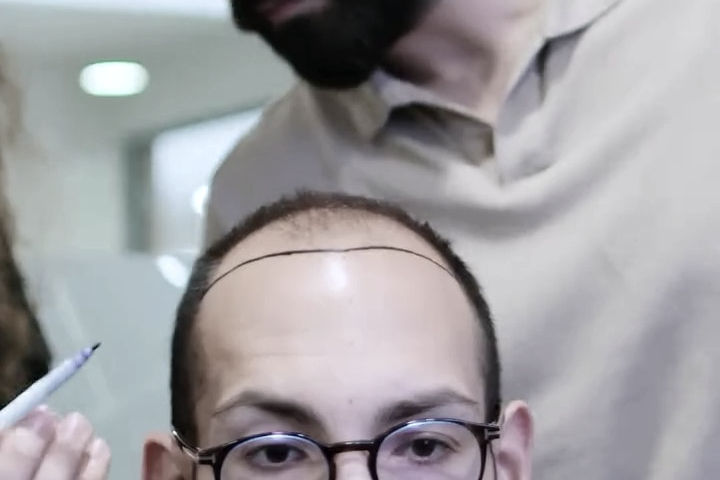
The Norwood Scale is an exceptional tool for geographical mapping, providing a common language for patients and doctors to discuss the visible progression of male pattern baldness. However, it is fundamentally a two-dimensional chart. It elegantly illustrates where the hair is missing, but it completely ignores the complex, three-dimensional biological realities that ultimately dictate whether a hair transplant will be successful—and how natural it will look.
Relying purely on a Norwood number to determine surgical candidacy is a clinical oversight. In practice, two patients can sit side-by-side in our clinic, both perfectly categorized as a Norwood Stage 4, yet present with completely different surgical viabilities and aesthetic outcomes.
Here are the critical, hidden biological variables that the Norwood Scale fails to capture:
1. Donor Density (Your Biological Bank Account)
The Norwood Scale measures the recipient area (the balding zone), but it ignores the donor area (the back and sides of the head). A hair transplant requires moving follicles from the donor zone to the balding areas.
A Stage 4 patient with an exceptionally dense donor zone might safely yield 4,000 grafts without the back of their head looking thin.
Conversely, a Stage 4 patient with naturally low donor density might only be able to safely yield 2,500 grafts.
This means that despite having the exact same Norwood classification, the first patient can achieve a thick, dense restoration across their entire scalp, while the second patient will require a highly conservative approach, prioritizing the hairline and leaving the crown lighter.
2. Hair Caliber (The Variable of Light and Opacity)
Hair caliber refers to the microscopic thickness (diameter) of your individual hair shafts. When it comes to achieving visual density, caliber is arguably more important than the raw number of grafts. A hair transplant works by creating an illusion of density by blocking light from reflecting off the scalp. Thicker hair shafts physically block more light.
Therefore, a Norwood 4 patient with thick, coarse hair might only need 2,500 grafts to achieve a full, opaque look.
A Norwood 4 patient with very fine, thin hair might need 3,500 grafts just to achieve the exact same visual density, simply because thinner hairs offer less coverage per graft.
3. Diffuse Miniaturization (The Invisible Threat)
The standard Norwood scale was designed to measure bare skin—the slick, completely bald areas of the scalp. What it struggles to account for is diffuse miniaturization. Many patients present with a distinct Norwood pattern (such as a Stage 3 hairline), but the hair sitting directly behind that hairline is actively miniaturizing and weakening. If a surgeon only focuses on restoring the bare "M" shape (as indicated by the Norwood scale) and ignores the weakening hair behind it, the transplant will look disconnected and unnatural within a few years as that native hair inevitably falls out.
The Eva Estetica Standard: This is precisely why a visual assessment or a submitted photograph is never enough to build a surgical plan. The Norwood Scale provides the outline, but it requires a high-definition digital trichoscopy audit to calculate the density, caliber, and true miniaturization boundaries that will fill in the details of your final restoration.
The Age Factor: The Trap of the 20-Something Norwood 3

The Norwood Scale measures the physical geography of your hair loss, but it completely lacks the context of time. From a surgical perspective, a patient’s age is arguably the most critical variable that the scale ignores.
A Norwood Stage 3 at age 25 and a Norwood Stage 3 at age 45 require entirely different clinical approaches.
The 25-Year-Old Norwood 3 (High Risk): If you have reached clinical baldness in your mid-20s, your genetic sensitivity to DHT is exceptionally aggressive. Your hair loss is moving fast. If a surgeon simply performs a transplant to fill in your receded temples without medically stabilizing your native hair first, you will continue to lose the hair behind the transplant. By age 30, you could be left with a transplanted hairline, a massive bald gap behind it, and a depleted donor area. For young patients, medical stabilization is a mandatory prerequisite to surgery.
The 45-Year-Old Norwood 3 (High Stability): If you have only reached a Stage 3 by your mid-40s, your hair loss is progressing very slowly. Your pattern is largely "locked in," and your donor area has proven its long-term genetic resilience. Patients in this category are exceptional surgical candidates because the clinical team can design a dense, mature hairline with near-absolute certainty that the underlying native hair will not aggressively vanish in the coming decade.
Understanding the relationship between your age and your Norwood stage is what separates a short-sighted cosmetic fix from a lifelong architectural triumph.
Conclusion: Moving from Diagnosis to Design
Identifying your place on the Norwood Scale is a vital starting point for your research. It gives a medical name to the pattern you see in the mirror and helps you communicate with clinical professionals. However, viewing your hair loss solely through the lens of a two-dimensional chart is a common clinical trap.
The most important educational takeaway is this: the Norwood Scale only captures a snapshot of your hair loss today, but androgenetic alopecia is a progressive, lifelong condition.
At Eva Estetica Istanbul, we go far beyond the Norwood Scale. Treating a "Norwood Stage" in isolation often leads to short-sighted surgical planning. If a clinic simply treats your current stage by packing thousands of grafts into your immediate bald spots—without calculating your future hair loss trajectory—you risk entirely depleting your finite donor reserve. If your hair loss progresses to the next Norwood stage five years later, you will have no donor hair left to fix it.
A truly sophisticated restoration strategy requires understanding that you have a limited "biological bank account" of grafts on the back of your head. They must be spent wisely to frame your face today, while holding enough in reserve for the stages you may reach a decade from now.
We do not treat a number; we treat the complete biological ecosystem of your specific scalp. A successful, undetectable hair transplant requires blending the geographical data of the Norwood map with the complex, three-dimensional reality of your hair caliber, underlying miniaturization, and individual facial geometry.
By conducting a high-definition digital trichoscopy audit, we map your invisible boundaries of thinning, transitioning from generalized estimates to exact surgical mathematics. Our ultimate commitment is to ensure that your surgical blueprint is not just medically safe for today, but architecturally designed to look completely natural, age-appropriate, and enduring for the rest of your life.
Frequently Asked Questions: The Norwood Scale
1. Is a Norwood 2 hairline considered balding?
No. A Norwood Stage 2 is typically a mature hairline, featuring a slight, symmetrical recession of 1 to 1.5 cm at the temples. This is a natural, biological part of facial aging in adult men, not an active state of clinical baldness, and ethical surgeons will usually refuse to operate on it.
2. Can I reverse a Norwood 3 with medication alone?
Medications like Finasteride and Minoxidil are highly effective at halting further progression and thickening miniaturizing hairs, particularly at the crown. However, they cannot regrow hair on completely slick, bare skin. To restore the deeply receded temporal peaks of a true Norwood 3, a surgical hair transplant is required.
3. At what Norwood stage is it too late for a hair transplant?
Norwood Stage 7 is generally considered the limit for surgical candidacy. Because the balding area on top of the head is incredibly vast and the remaining donor hair on the sides is very thin, a full restoration is biologically impossible. While minor, ultra-conservative coverage can sometimes be achieved, expectations must be heavily managed.
4. How fast will I progress from a Norwood 3 to a Norwood 4?
Hair loss progression is entirely unpredictable and non-linear. Instead of a steady decline, patients often experience periods of apparent stability followed by aggressive bursts of thinning. While a specific timeline cannot be predicted, the trajectory will only worsen without medical intervention to block DHT.
5. Can I get 6,000 grafts in one session for a Norwood 5 or 6?
While advanced Norwood stages require a high volume of grafts, ethical clinics strictly advise against "mega-sessions" (extracting 6,000+ grafts in a single day). Pushing beyond the scalp's biological limits in one sitting massively increases the risk of donor depletion, vascular necrosis, and graft failure. It is much safer to stage the restoration across two separate surgeries.
6. What is a Norwood Class A hair loss pattern?
Unlike the standard progression that creates an "M" shape at the front and an isolated bald spot on the crown, a Norwood Class A progression involves the entire frontal hairline receding uniformly backward in a relatively straight line, with the crown remaining stable early on.
7. Should I wait until I am a Norwood 5 to get a hair transplant?
No, waiting is mathematically disadvantageous. The ideal time for surgical intervention is often between Norwood Stage 3 and Stage 4. If you wait until Stage 5 or 6, the balding area becomes vastly larger while your available donor supply becomes smaller, making full density much harder to achieve.
8. What is the difference between the Norwood Scale and the Ludwig Scale?
The Norwood Scale maps male pattern baldness, which is characterized by geometric recession and distinct bald spots. The Ludwig Scale maps female pattern hair loss, which typically presents as widespread, diffuse thinning across the entire top of the scalp while the original frontal hairline remains firmly intact.
Academic Sources and Clinical Literature
The staging systems, diagnostic protocols, and medical therapies discussed in this guide are deeply rooted in foundational dermatological literature and peer-reviewed medical studies. For further reading on the clinical development of the Norwood scale and the realities of modern hair restoration, please consult the following texts:
1. Male Pattern Baldness: Classification and Incidence Norwood OT. Southern Medical Journal (1975). This is the universally recognized foundational paper that established the modern Norwood Scale. Dr. O'Tar Norwood revised previous classification systems to create the definitive medical standard used by surgeons globally today to categorize the progressive stages of male pattern hair loss.
View on PubMed: https://pubmed.ncbi.nlm.nih.gov/1188424/
2. Classification of Male-pattern Hair Loss Wirya CT, Wu W, Wu K. International Journal of Trichology (2017). A comprehensive, peer-reviewed clinical update evaluating the history, detail, practicality, and reproducibility of the Norwood-Hamilton classification system. This paper is essential for understanding how medical professionals universally diagnose the severity and progression of androgenetic alopecia today.
View on PubMed Central: https://pmc.ncbi.nlm.nih.gov/articles/PMC5596658/
3. Effect of Follicular Unit Extraction on the Donor Area Mohmand MH, Ahmad M. World Journal of Plastic Surgery (2018). A critical clinical study evaluating the exact mathematical impact of FUE extraction on a patient's donor reserve. The research demonstrates that extracting a high percentage of follicular units significantly decreases local hair mass and visual density. The study concludes that because native donor density varies so drastically between patients, high-volume FUE must be performed with extreme medical caution to prevent permanent depletion.
View on PubMed Central: https://pmc.ncbi.nlm.nih.gov/articles/PMC6066700/


Comments